The science of engineering fluid streams at the microscale, known as microfluidics, offers several technological advantages when applied to biomedical research. One of its abilities is to reproduce biological activities and different biological functions while better emulating the in vivo settings. With microfluidics it is possible not only to decrease volumes, reagents, and experimental times, but also to increase the precise control of the microenvironment. Our laboratory is involved in several projects that exploit this technology applied on cancer studies.
Cancer cell migration studies
The study of tumor cell migration is of paramount importance in biological and medical research, as it is a key event in cancer metastasis. Metastasis is the dynamical process in which cancer cells begin to move in the surrounding microenvironment, driven by mainly different chemical and mechanical stimuli. Initial stage migration information with single-cell resolution may play an exclusive role in metastasis. Consequently, the need for a device that is able to study cancer migration at this scale and resolution becomes crucial. Applying an unprecedented fabrication strategy that combines two-photon-polymerization and lithography, a single-cell migration platform was developed.
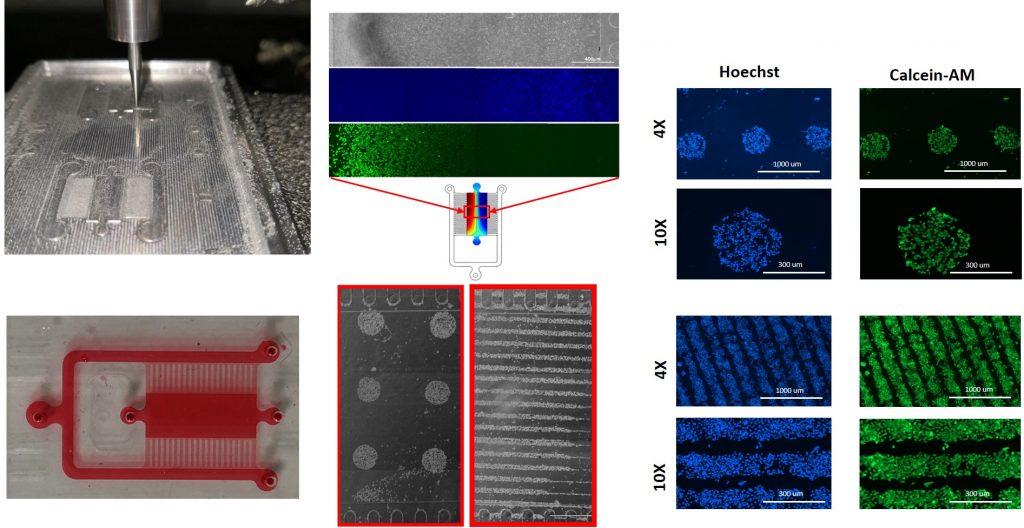
Patterning of cancer cells inside an Extracellular Vesicles (EVs) microgradient generating device
Cell patterning in an hydraulically-sealed microfluidic device remains challenging. This method was used to create cell-adhesive and non-adhesive areas on a glass substrate. The patterned substrate was used for selective attachment of cancer cells and was combined with the microfluidic platform. This gradient-generating microfluidic platform could be useful to assess exosomes-induced concentration-dependent cellular responses. In recent years, Extracellular Vesicles (EVs) are being recognized as contributing factors in mediating cancer progression. Due to the relatively short history of EVs in the literature, there are only a few studies employing microfluidic platforms for studying their role in cancer metastasis. Microfluidic approaches for the generation of controllable and stable concentration gradients coupled with the ability to micropattern the cell culture surface area could enable fast screenings of induced concentration-dependent signals and could surpass several limitations of conventional cell culture techniques.
Neuroblastoma on a chip platform
Despite great progresses achieved in the understanding of cancer biology, metastases are still synonymous of terminal illness in several cancers, including Neuroblastoma (NB), an embryonal malignancy of early childhood. Recent studies highlighted the importance of the complex multicellular, biochemical, and biophysical stimuli regulating tumor behavior during progression and metastasis. Microscaled devices specific organ-on-a-chip platforms can reproduce such tailored microenvironments and provide more relevant in vitro models for drug screening applications and for a better understanding of tumor metastatic dissemination. The creation of cost-effective and reliable in vitro models that can be used for accurately screening anticancer drug effects as well as overcoming the drawbacks of conventional models is of great importance for improving the clinical management of primary tumor and metastatic cancers.
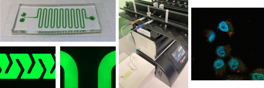
Microfluidic devices for EVs engineering
Despite being the guilty part of cancer progression, extracellular vesicles (EVs) could potentially play a role in the treatment of cancers by transforming them into drug delivery systems. Although traditional methods for EVs modifications will continue to play a significant role in the future, it is believed that microfluidic approaches will eventually replace benchtop methods for engineering EVs. Considering such promises, the goal of the project is to develop a microfluidic device for generating drug-loaded EVs. EVs can be loaded with various therapeutic agents, including Verteporfin (VP). VP is a porphyrin that has recently provided positive results for its anti-cancer activity in Neuroblastoma – one of the most common solid tumors of childhood. Taking advantage of lab-on-chip devices, passive VP-loading through microfluidic mixing and incubation may be an important strategy to produce functional engineered EVs. The platform is designed using AutoCAD® and modeled with COMSOL Multiphysics ® so that both a perfect mixing between EVs and VP occurs within the mixing unit and the desired incubation time is achieved. The master is fabricated with photolithography, and replicas of the chips are obtained with replica molding processes. Plasma treatment is used to form a hydraulic seal of the platform to a glass support. The device in this configuration is used to perform both fluid dynamic and biological validations, confirming that highly efficient loading of VP into EVs is obtained. We are confident that microfluidic technology, with its versatility and precision, could fully unlock the capability of EVs for speeding up precision therapeutics.